Jemma Kann
  • Blog
  • About
  • Contact

The Treatment Of Over-Pronation

6/6/2015

 
Overview

Over pronation is when there is excessive or too much movement of the foot. Over pronation is a very common cause of heel pain and general pain throughout the lower extremities. This condition can often be referred to as flat feet and causes you to walk on other parts of your foot, which is what leads to serious heel and foot pain.Overpronation

Causes

There are many possible causes for overpronation, but researchers have not yet determined one underlying cause. Hintermann states, Compensatory overpronation may occur for anatomical reasons, such as a tibia vara of 10 degrees or more, forefoot varus, leg length discrepancy, ligamentous laxity, or because of muscular weakness or tightness in the gastrocnemius and soleus muscles. Pronation can be influenced by sources outside of the body as well. Shoes have been shown to significantly influence pronation. Hintermann states that the same person can have different amounts of pronation just by using different running shoes. It is easily possible that the maximal ankle joint eversion movement is 31 degrees for one and 12 degrees for another running shoe.

Symptoms

It is important to note that pronation is not wrong or bad for you. In fact, our feet need to pronate and supinate to achieve proper gait. Pronation (rolling inwards) absorbs shock and supination (rolling outwards) propels our feet forward. It is our body?s natural shock-absorbing mechanism. The problem is over-pronation i.e. the pronation movement goes too deep and lasts for too long, which hinders the foot from recovering and supinating. With every step, excess pronation impedes your natural walking pattern, causing an imbalance in the body and consequent excessive wear and tear in joints, muscles and ligaments. Some common complaints associated with over-pronation include Heel Pain (Plantar Fasciitis) ,Ball of foot pain, Achilles Tendonitis, Shin splints, Knee Pain, Lower Back Pain.

Diagnosis

Look at the wear on your shoes and especially running trainers; if you overpronate it's likely the inside of your shoe will be worn down (or seem crushed if they're soft shoes) from the extra strain.Over-Pronation

Non Surgical Treatment

Supportive orthotics in the shoe is a method commonly implemented to treat many common running injuries associated with pronation. An advantage of orthotics is that they often allow the sufferer to continue to participate in athletic activity and avoid other treatment options that could be potentially costly and time consuming. Seventy-five percent of injured runners are successfully treated with the prescription of orthoses. Orthotics are the most effective treatment for symptoms that develop from unusual biomechanics within the body such as overpronation, resulting in either great improvement or complete healing of the injury in about half the cases.

Prevention

Many of the prevention methods for overpronation-orthotics, for example-can be used interchangeably with treatment methods. If the overpronation is severe, you should seek medical attention from a podiatrist who can cast you for custom-made orthotics. Custom-made orthotics are more expensive, but they last longer and provide support, stability, and balance for the entire foot. You can also talk with a shoe specialist about running shoes that offer extra medial support and firm heel counters. Proper shoes can improve symptoms quickly and prevent them from recurring. Surgery can sometimes help cure and prevent this problem if you suffer from inherited or acquired pes planus deformity. Surgery typically involves stabilizing the bones to improve the foot?s support and function.

Calcaneal Apophysitis Physical Therapy

5/20/2015

 
Overview

Sever disease, first described in 1912, is a painful inflammation of the calcaneal apophysis. It is classified with the child and adolescent nonarticular osteochondroses. (The other disease in this group is Iselin disease, which is inflammation of the base of the fifth metatarsal.) The etiology of pain in Sever disease is believed to be repetitive trauma to the weaker structure of the apophysis, induced by the pull of the tendo calcaneus (Achilles tendon) on its insertion. This results in a clinical picture of heel pain in a growing active child, which worsens with activity. Sever disease is a self-limited condition; accordingly, no known complication exists from failure to make the correct diagnosis.

Causes

Sever's Disease typically affects boys and girls between 8-15 years of age. Risk factors include. Athletic activity that involves heel contact with hard surfaces, as in gymnastics, track, soccer, basketball, ice skating, ballet and aerobics. The wearing of ill-fitting shoes. Well-made shoes that fit properly are a must for every child. Prolonged periods of standing. If a child complains of heel pain after choir practice, doing dishes, standing in lines or other activities that put pressure on the heel bones, pay attention.

Symptoms

Athletes with Sever?s disease are typically aged 9 to 13 years and participate in running or jumping sports such as soccer, football, basketball, baseball, and gymnastics. The typical complaint is heel pain that develops slowly and occurs with activity. The pain is usually described like a bruise. There is rarely swelling or visible bruising. The pain is usually worse with running in cleats or shoes that have limited heel lift, cushion, and arch support. The pain usually goes away with rest and rarely occurs with low-impact sports such as bicycling, skating, or swimming.

Diagnosis

Radiography. Most of the time radiographs are not helpful because the calcaneal apophysis is frequently fragmented and dense in normal children. But they can be used to exclude other traumas. Ultrasonography. could show the fragmentation of secondary nucleus of ossification of the calcaneus in severs?s disease. This is a safe diagnostic tool since there is no radiation. This diagnostic tool can also be used to exclude Achilles tendinitis and/or retrocalcaneal bursitis.

Non Surgical Treatment

Occasionally, an orthotic may need to be prescribed for temporary or long-term correction of their foot biomechanics (eg flat feet or high arches). During the acute phase of Sever's disease a small heel rise or shock-absorbing heel cup placed under the heel pad of your child's foot may help to ease the symptoms. Your podiatrist or physiotherapist can assess your child's arch and guide you in the best management of your child's condition. We recommend that your child should never go barefooted during the painful stages of Sever's disease.

Foot Pain In The Arch

4/16/2015

 
Overview

One of those often-painful soft tissue that attaches to heel spurs at the bottom of the foot is called "plantar fascia". Fascia, located throughout the body, is a fibrous connective tissue similar to a ligament. You can see fascia when you handle meat. It is the white, connective tissue separating layers of meat or attaching to bones. The "plantar" fascia in our bodies is that fascia which is seen on the bottom (or plantar portion) of the foot, extending from the heel bone to the ball of the foot. Compared to other fascia around the body, plantar fascia is very thick and very strong. It has to be strong because of the tremendous amount of force it must endure when you walk, run or jump. But while the plantar fascia is a strong structure, it can still get injured, most commonly when it is stretched beyond its normal length over long periods of time. When plantar fascia is injured, the condition is called "plantar fasciitis", which is usualy pronounced either "plan-tar fash-I-tis" or "plan-tar-fash-ee-I-tis." (Adding "-itis" to the end of a word means that structure is inflamed.) It is sometimes known more simply as 'fasciitis'. Plantar fasciitis is the most common type of arch pain.

Pain In Arch

Causes

Flat feet are often hereditary. Arch pain may also be caused by wearing shoes with inadequate support, standing or walking for long periods of time in high heels, or overuse of the feet during work or sports. Being overweight also places additional stress on the feet, especially the arches.

Symptoms

Symptoms include pain which is often described as a burning pain radiating into the arch of the foot, heel and sometimes the toes. Pins and needles or numbness may be felt in the sole of the foot. Pain may be worse when running or when standing for long periods of time and often worse at night. The area under the medial malleolus on the inside of the ankle may be tender to touch.

Diagnosis

Your doctor may order imaging tests to help make sure your heel pain is caused by plantar fasciitis and not another problem. X-rays provide clear images of bones. They are useful in ruling out other causes of heel pain, such as fractures or arthritis. Heel spurs can be seen on an x-ray. Other imaging tests, such as magnetic resonance imaging (MRI) and ultrasound, are not routinely used to diagnose plantar fasciitis. They are rarely ordered. An MRI scan may be used if the heel pain is not relieved by initial treatment methods.

Non Surgical Treatment

The most effective treatment for foot arch pain and strain is to use an arch support. The arch support sits under the foot and stops the arch of the foot from collapsing, thereby preventing the stretch of the arch pad which causes pain and discomfort. Wearing an arch support in slippers or house shoes can also prevent pain in the mornings when discomfort it most common and severe. Arch supports usually relieve symptoms within a few days.

Pain In Arch

Surgical Treatment

The soft tissue surgeries usually would include a lengthening of the Achilles tendon, releasing of the plantar fascia as well as tendon transfers. These procedures are usually done in conjunction with bony procedures such as calcaneal osteotomies (to lower the heel bone and get it more under the leg itself), as well as metatarsal osteotomies. These procedures usually involve either cutting or fusion of the bones, and placement of fixation devices to allow the bones to heal. Healing time is usually at least 6-8 weeks and usually the patient must be non-weight bearing during the healing process. These types of surgical corrections are usually reserved for the more difficult, painful and deformed feet. They can require more surgeries down the line. These procedures are usually the last resort after all other modes of treatment have been exhausted (except in children where it is usually best to treat the deformity early). There are many different degrees of high arched feet and these procedures should be left for the more extreme cases. These cases usually require a very high degree of surgical skill and should only be done by those who frequently perform these types of cases.

Prevention

Foot and ankle injuries are common in sports, especially running, tennis and soccer. But sports enthusiasts can decrease the risk of injury by taking some precautions. Lightly stretch or better yet, do a slow jog for two to three minutes to warm up the muscles. Don't force the stretch with a "bouncing motion." The amount of time spent on the activity should be increased gradually over a period of weeks to build both muscle strength and mobility. Cross training by participating in different activities can help build the muscles. People whose feet pronate or who have low arches should choose shoes that provide support in both the front of the shoe and under the arch. The heel and heel counter (back of the shoe) should be very stable. Those with a stiffer foot or high arches should choose shoes with more cushion and a softer platform. Use sport-specific shoes. Cross training shoes are an overall good choice; however, it is best to use shoes designed for the sport.

Stretching Exercises

Gastroc stretch. Stand on the edge of a step. Rise slowly on your toes. Lower yourself slowly as far as you can until you feel a stretch in your calf. Don?t roll your foot inward or outward. Hold for 1-2 seconds. Reps:10-20 (stop before you fatigue). Soleus stretch. Same as above, but start with your knee bent so that you feel a slight stretch in your calf or achilles. Maintain the angle of your knee throughout the stretch. Bicycle stretch. Lie on your side. Keeping your top leg straight, bring your knee toward your nose until you feel a slight stretch in the hamstring. Maintaining this angle at your hip, start pretending you are pedalling a bicycle with the top leg. Make sure you feel a slight stretch each time your knee is straight. Reps: 10-30 for each leg. If you feel any pops or clicks in your hip or back, try raising the top leg a little (making the thighs further apart) to eliminate the popping. Foot Intrinsic Exercises. Assisted metatarsal head raising. Sit in a chair. Find the bumps at the ball of your foot just before your big toe and just before the little toe. These are the first (big toe) and fifth (little toe) metatarsal heads. Place your second and third fingers from one hand under the first metatarsal head, and the second and third fingers from the other hand under the fifth metatarsal head. Now lay the thumbs from each hand in a diagonal across your toes so that they form a right angle meeting at the nail of the second toe. Your hands are now in position to assist your toes. Keep your toes straight, with the toe pads on the floor. Use your fingers to help raise all the metatarsal heads (the ball of your foot). Do not let your toes curl under keep them long. Now relax. Reps 7-10 for each foot. As this exercise gets easier, let your fingers do less of the work until your toes can do the exercise unassisted. This can take up to three weeks. When your strength has improved to this point, you can progress to the following three exercises, which are best done in stocking feet on a slippery floor. Active metatarsal head raising. Stand with your weight on both feet. Raise your metatarsal heads (the ball of your foot) while keeping your toes from curling under and maintaining your heel on the ground. Relax. Reps 6-7. Do one foot at a time. If you do more reps than you are ready for, you may well develop cramping in your foot. I once had a client who thought if seven reps were good, 10 were better. For good measure, she did the 10 reps 10 times in a day, and then she was unable to walk the next day from having used a set of muscles she had never exercised before. Don?t overdo it.

Acute Achilles Tendon Rupture Surgery Rehab

4/13/2015

 
Overview Achilles Tendonitis A rupture of the Achilles tendon means that there has been either a complete, or partial, tear of the tendon which connects the calf muscles to the heel bone. Usually this occurs just above insertion on the heel bone, although it can happen anywhere along the course of the tendon. Causes Your Achilles tendon helps you point your foot downward, rise on your toes and push off your foot as you walk. You rely on it virtually every time you move your foot. Rupture usually occurs in the section of the tendon located within 2 1/2 inches (about 6 centimeters) of the point where it attaches to the heel bone. This section may be predisposed to rupture because it gets less blood flow, which also may impair its ability to heal. Ruptures often are caused by a sudden increase in the amount of stress on your Achilles tendon. Common examples include increasing the intensity of sports participation, especially in sports that involve jumping, falling from a height, stepping into a hole. Symptoms A classic sign of an Achilles tendon rupture is the feeling of being hit in the Achilles are. There is often a "pop" sound. There may be little pain, but the person can not lift up onto his toes while weight bearing. Diagnosis The diagnosis of an Achilles tendon rupture can be made easily by an orthopedic surgeon. The defect in the tendon is easy to see and to palpate. No x-ray, MRI or other tests are necessary. Non Surgical Treatment Initial treatment for sprains and strains should occur as soon as possible. Remember RICE! Rest the injured part. Pain is the body's signal to not move an injury. Ice the injury. This will limit the swelling and help with the spasm. Compress the injured area. This again, limits the swelling. Be careful not to apply a wrap so tightly that it might act as a tourniquet and cut off the blood supply. Elevate the injured part. This lets gravity help reduce the swelling by allowing fluid and blood to drain downhill to the heart. Over-the-counter pain medication is an option. Acetaminophen (Tylenol) is helpful for pain, but ibuprofen (Motrin, Advil, Nuprin) might be better, because these medications relieve both pain and inflammation. Remember to follow the guidelines on the bottle for appropriate amounts of medicine, especially for children and teens. Achilles Tendinitis Surgical Treatment Surgical techniques for rupture repair are varied but usually involve reapproximation of the torn ends of the Achilles tendon, sometimes reinforced by the gastrocsoleus aponeurosis or plantaris tendon. Open reconstruction is undertaken using a medial longitudinal approach. Studies indicate that patients who undergo percutaneous, rather than an open, Achilles tendon rupture repair have a minimal rate of infection but a high rate of sural nerve entrapment (16.7% of treated cases). Prevention Good flexibility of the calf muscles plays an essential role in the prevention of Achilles tendon injuries. It is also important to include balance and stability work as part of the training programme. This should include work for the deep-seated abdominal muscles and for the muscles that control the hip. This might at first appear odd, given the fact that the Achilles are a good distance from these areas, but developing strength and control in this area (core stability) can boost control at the knee and ankle joints. Training errors should be avoided. The volume, intensity and frequency of training should be monitored carefully, and gradually progressed, particularly when introducing new modes of training to the programme. Abrupt changes in training load are the primary cause of Achilles tendinopathy.

Achilles Tendonitis

4/4/2015

 
Overview

Achilles Tendinitis
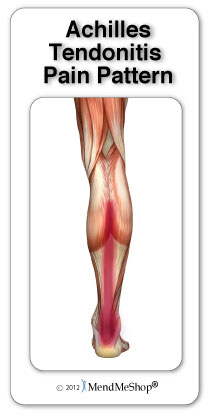
Do you experience dull pain near the back of your heel or in the back of your leg after your regular run or after playing your favourite sport? When you ramp up your exercise is the pain more severe or prolonged? If so, you may have Achilles tendinitis. The Achilles tendon is the thick, strong, springy band of tissue that connects the muscles from the middle of your calf to your heel bone. You use your Achilles tendon when you walk, run or jump. Achilles tendinitis occurs when the Achilles tendon is repeatedly strained. The Achilles tendon becomes less flexible, weaker and more prone to injury as we age. Middle-aged weekend warriors and runners who suddenly intensify their training often suffer from Achilles tendinitis.

Causes

There are hundreds of tendons scattered throughout our body, but it tends to be a small handful of specific tendons that cause problems. These tendons usually have an area of poor blood supply that leads to tissue damage and poor healing response. This area of a tendon that is prone to injury is called a "watershed zone," an area when the blood supply to the tendon is weakest. In these watershed zones, they body has a hard time delivering oxygen and nutrients necessary for tendon healing, that's why we see common tendon problems in the same parts of the body. Tendonitis is most often an overuse injury. Often people begin a new activity or exercise that causes the tendon to become irritated. Tendon problems are most common in the 40-60 year old age range. Tendons are not as elastic and forgiving as in younger individuals, yet bodies are still exerting with the same force. Occasionally, there is an anatomical cause for tendonitis. If the tendon does not have a smooth path to glide along, it will be more likely to become irritated and inflamed. In these unusual situations, surgical treatment may be necessary to realign the tendon.

Symptoms

Achilles tendonitis may be felt as a burning pain at the beginning of activity, which gets less during activity and then worsens following activity. The tendon may feel stiff first thing in the morning or at the beginning of exercise. Achilles tendonitis usually causes pain, stiffness, and loss of strength in the affected area. The pain may get worse when you use your Achilles tendon. You may have more pain and stiffness during the night or when you get up in the morning. The area may be tender, red, warm, or swollen if there is inflammation. You may notice a crunchy sound or feeling when you use the tendon.

Diagnosis

A doctor examines the patient, checking for pain and swelling along the posterior of the leg. The doctor interviews the patient regarding the onset, history, and description of pain and weakness. The muscles, tissues, bones, and blood vessels may be evaluated with imaging studies, such as X-ray, ultrasound, or MRI.

Nonsurgical Treatment

Conservative management of Achilles tendinosis and paratenonitis includes the following. Physical therapy. Eccentric exercises are the cornerstone of strengthening treatment, with most patients achieving 60-90% pain relief. Orthotic therapy in Achilles tendinosis consists of the use of heel lifts. Nonsteroidal anti-inflammatory drugs (NSAIDs): Tendinosis tends to be less responsive than paratenonitis to NSAIDs. Steroid injections. Although these provide short-term relief of painful symptoms, there is concern that they can weaken the tendon, leading to rupture. Vessel sclerosis. Platelet-rich plasma injections. Nitric oxide. Shock-wave therapy. Surgery may also be used in the treatment of Achilles tendinosis and paratenonitis. In paratenonitis, fibrotic adhesions and nodules are excised, freeing up the tendon. Longitudinal tenotomies may be performed to decompress the tendon. Satisfactory results have been obtained in 75-100% of cases. In tendinosis, in addition to the above procedures, the degenerated portions of the tendon and any osteophytes are excised. Haglund?s deformity, if present, is removed. If the remaining tendon is too thin and weak, the plantaris or flexor hallucis longus tendon can be weaved through the Achilles tendon to provide more strength. The outcome is generally less favorable than it is in paratenonitis surgery.

Achilles Tendon

Surgical Treatment

Percutaneous Achilles Tendon Surgery. During this procedure the surgeon will make 3 to 4 incisions (approx. 2.5 cm long) on both sides of the Achilles tendon. Small forceps are used to free the tendon sheath (the soft tissue casing around your Achilles tendon) to make room for the surgeon to stitch/suture any tears. Skilled surgeons may perform a percutaneous achilles tendon surgery with ultrasound imaging techniques to allow for blink suturing with stab incisions made by a surgical suture needle. This procedure can be done in 3 different ways depending on the preference and experience of your surgeon. Instead of making several 2.5 cm incisions for this procedure, some surgeons will use guided imaging with an ultrasound to see the Achilles tendon tissue without having to open up your ankle. For this technique, they will use a surgical needle to repeatedly stab your Achilles tendon. These "stab incisions" will allow the surgeon to "blindly" suture your tendon without seeing the actual tissue. As another option - some surgeons will only make 1 to 3 incisions for smaller surgical implements to repair your tendon while relying on imaging ultrasound to see your damaged tissue. During either procedure the use of ultrasound imaging or endoscopic techniques requires a very skilled surgeon.

Prevention

A 2014 study looked at the effect of using foot orthotics on the Achilles tendon. The researchers found that running with foot orthotics resulted in a significant decrease in Achilles tendon load compared to running without orthotics. This study indicates that foot orthoses may act to reduce the incidence of chronic Achilles tendon pathologies in runners by reducing stress on the Achilles tendon1. Orthotics seem to reduce load on the Achilles tendon by reducing excessive pronation,

What Is Pes Planus?

4/1/2015

 
Overview

Adult Acquired Flat Foot

Flat feet is the most common foot deformity known. In fact, sixty million Americans or 25% of the U.S. population have flat feet. Some of these people may experience problems that limit their activities, while others can run marathons or play in the NBA. If you've ever seen your footprints in the sand and they looked more like bricks than feet, then you probably have flat feet. Simply stated, a flat foot is a foot that does not have an arch when standing. In the medical world, flat feet are associated with "pronated" feet. Pronated is merely the term used to describe the position of the foot when it is flexed upward (dorsiflexed), turned away from the body (abducted), and the heel is rolled outward (everted), all at the same time. A certain amount of pronation is required for normal walking, but too much pronation is often considered a foot's "worst enemy." Over time, excessive pronation can lead to many unpleasant problems including heel pain, bunions, hammertoes, shin splints, and even knee, hip, or back pain. In fact, one orthopedic surgeon discovered that 95% of his total knee replacement patients and 90% of his total hip replacement patients had flat feet. An easy way to tell if you pronate too much is to take a look at your athletic shoes-excessive wearing of the inside heel (arch side of the shoe) as compared to the outside is a classic indication of excessive pronation.


Causes

When flat feet develop at a later age, they are known as fallen arches. The arches may fall because the muscles supporting them are no longer able to do so. In addition the spring ligament within the foot may have lost some of its tension allowing the arch of the foot to flatten. Other conditions causing fallen arches include sudden weight gain, a nervous system injury, or a loss of sensation caused by a disease such as diabetes. Most people with fallen arches are flat on both feet.


Symptoms

Knee/Hip/Back Pain - When the arch collapses in the foot, it triggers a series of compensations up the joint chain, leading to increased stress on the knee, pelvis and low back. Plantar fasciitis - This condition is characterized by heel pain, especially with the first few steps you take. The plantar fascia stretches as the arch falls, putting stress on the heel. Bunions - If you see a bony bump developing at the base of your big toe, you are likely developing a bunion. It may be swollen, red or painful when it rubs against your shoe. A flattened arch spreads the forefoot and causes the big toe to deviate toward the second toe. Shin splints - This term generally refers to pain anywhere along the shinbone. It is typically due to overuse and is aggravated after exercise and activity.


Diagnosis

An examination of the foot is enough for the health care provider to diagnose flat foot. However, the cause must be determined. If an arch develops when the patient stands on his or her toes, the flat foot is called flexible and no treatment or further work-up is necessary. If there is pain associated with the foot or if the arch does not develop with toe-standing, x-rays are necessary. If a tarsal coalition is suspected, a CT scan is often ordered. If a posterior tibial tendon injury is suspected, your health care provider may recommend an MRI.


Non Surgical Treatment

Most cases of flatfeet do not require treatment. However, if there is pain, or if the condition is caused by something other than normal development, there are several treatment options. Self-care options include rest, choosing non-weight-bearing exercise (e.g., swimming, cycling), weight loss, and avoiding high heels. Flexible flatfeet with some pain can be relieved with the use of orthotics-shoe inserts that support the arch-and/or heel wedges (in some cases). If pronation is a factor, special shoes can be worn that lift the arch and correct the inward leaning. Physical therapy may also be prescribed to stretch or lengthen the heel cord and other tendons. For rigid or inflexible flatfeet, treatment varies depending on the cause. Tarsal coalition if often treated with rest and the wearing of a cast. If this is ineffective, surgery can be done to separate the bones or to reset the bones into a correct position. If the flatfoot is caused by an injury to the tendons in the foot or ankle, rest, anti-inflammatory medications (e.g., ibuprofen), and the use of shoe inserts and ankle braces often relieve symptoms. In severe cases, surgery is performed to repair the tendon or to fuse some joints in the foot into a corrected position to reduce stress on the tendon. The prognosis after surgery is generally good. Complications include pain and some loss of ankle motion, especially when trying to turn the foot in or out. This may be improved with physical therapy.


Surgical Treatment

Acquired Flat Foot

Common indications for surgery are cerebral palsy with an equinovalgus foot, to prevent progression and breakdown of the midfoot. Rigid and painful Pes Planus. To prevent progression, eg with a Charcot joint. Tibialis posterior dysfunction, where non-surgical treatment is unsuccessful. Possible surgical procedures include Achilles tendon lengthening. Calcaneal osteotomy, to re-align the hindfoot. Reconstruction of the tibialis posterior tendon. For severe midfoot collapse of the arch, triple arthrodesis may be indicated.

Pain In The Arches Cause And Effect

3/27/2015

 
Overview

Plantar fasciitis continues to be widely used for the clinical syndrome of undersurface heel pain. However, the use of 'itis' denotes an inflammatory disorder. This is a misnomer as the pathology is not the result of excessive inflammation. Pathological changes are degenerative (but partially reversible) in nature, probably due to repetitive trauma. The plantar fascia is a thick, fibrous band of connective tissue. Its origin is the medial plantar tubercle of the calcaneum. It runs along the sole of the foot like a fan, being attached at its other end to the base of each of the toes. It is a tough, resilient structure that has a number of critical functions during running and walking. It stabilises the metatarsal joints during impact with the ground. It acts as a shock absorber for the entire leg. It forms the longitudinal arch of the foot and helps to lift the arch to prepare it for the 'take-off' phase of the gait cycle.

Arch Pain

Causes

If the feet are exposed to damp conditions for prolonged periods, you can develop Trench Foot. Most commonly associated with World War One, it now tends to be seen in builders, hikers or festival goers. Trench foot causing numbness and bottom of foot pain. With Trench Foot, the sole of the foot turns a white/grey colour and you may develop pins and needles or numbness. Other symptoms include pain in bottom of foot and swelling. If left untreated, you can develop blisters and permanent nerve damage which can lead to the need for amputation. Treatment and prevention aim to reduce the dampness around the foot and ensure good foot hygiene. Slowly rewarming the feet and using special products in a foot bath really helps reduce the damage and foot arch pain from Trench Foot.

Symptoms

Flat feet don't usually cause problems, but they can put a strain on your muscles and ligaments (ligaments link two bones together at a joint). This may cause pain in your legs when you walk. If you have flat feet, you may experience pain in any of the following areas, the inside of your ankle, the arch of your foot, the outer side of your foot, the calf, the knee, hip or back, Some people with flat feet find that their weight is distributed unevenly, particularly if their foot rolls inwards too much (overpronates). If your foot overpronates, your shoes are likely to wear out quickly. Overpronation can also damage your ankle joint and Achilles tendon (the large tendon at the back of your ankle). See your GP if you or your child has flat feet and your feet are painful, even when wearing supportive, well-fitting shoes, shoes wear out very quickly, feet appear to be getting flatter, feet are weak, numb or stiff, Your GP may refer you to a podiatrist (foot specialist).

Diagnosis

The doctor will examine your feet for foot flexibility and range of motion and feel for any tenderness or bony abnormalities. Depending on the results of this physical examination, foot X-rays may be recommended. X-rays are always performed in a young child with rigid flatfeet and in an adult with acquired flatfeet due to trauma.

Non Surgical Treatment

Treatment for a high arch foot or Charcot Marie Tooth disorder depends on the extent of deformity and the amount of disability experienced by the patient. Depending upon the symptoms, treatment may include. Changing the shoes. Special orthotic supports (devices that support, adjust, or accommodate the foot deformity). Cushioning pads. Foot and ankle braces or surgery.

Pain In Arch

Surgical Treatment

A procedure that involves placing a metallic implant (most commonly) at the junction where the foot meets the ankle. This device causes the physical blockade that prevent the collapse. It is a procedure that is only indicated for mobile feet, and should not be used with rigid flat feet. Dr. Blitz finds this procedure better for younger patients with flexible flat feet where the bone alignment is still developing so that the foot can adapt to function in a better aligned position.

Prevention

Foot and ankle injuries are common in sports, especially running, tennis and soccer. But sports enthusiasts can decrease the risk of injury by taking some precautions. Lightly stretch or better yet, do a slow jog for two to three minutes to warm up the muscles. Don't force the stretch with a "bouncing motion." The amount of time spent on the activity should be increased gradually over a period of weeks to build both muscle strength and mobility. Cross training by participating in different activities can help build the muscles. People whose feet pronate or who have low arches should choose shoes that provide support in both the front of the shoe and under the arch. The heel and heel counter (back of the shoe) should be very stable. Those with a stiffer foot or high arches should choose shoes with more cushion and a softer platform. Use sport-specific shoes. Cross training shoes are an overall good choice; however, it is best to use shoes designed for the sport.

Stretching Exercises

Ankle evert or strengthening. Lie on your side with your feet hanging off the end of your bed or a weight bench. Bend the toes of the foot that is closer to the ceiling slightly toward your head. This is the starting position. Now raise your toes toward the ceiling while keeping the rest of your leg stationary. Return to the starting position. Reps. 10-15. Now point your toes slightly away from your head. This is the starting position. Raise your toes toward the ceiling. Return to the starting position. Reps. 10-15. Ankle invertor strengthening. Same as above, but do the exercises with the foot that is closer to the floor. Dorsiflexor strengthening. Sit on a desk, table, or counter so that your feet don?t touch the ground. Let your feet dangle comfortably. Bend your foot upward as far as you can comfortably go. Do not let your foot pull inward or outward. Return to the starting position. Reps. 10-15.

Achilles Tendonitis

3/26/2015

 
Overview
Achilles Tendonitis The Achilles tendon is the extension from the two large muscles in the calf region, the gastrocnemius and the soleus. These two muscles combine to form the Achilles tendon. The tendon forms in the lower one third of the leg and extends to the back of the heel bone (calcaneus). When the muscles of the calf contract this produces tension on the Achilles tendon pulling on the back of heel causing the heel to rise and the foot to point downward. It is during this motion that high-tension force is transmitted through the Achilles tendon during pushing and jumping activity. This high tension force can cause the Achilles tendon to tear or rupture. This happens in 3 common locations. The most common location for a tendon tear is within the tendon substance just above the heel. The second and third most common locations are where the Achilles tendon attaches into the heel bone and higher in the leg, where the tendon begins.

Causes
An Achilles tendon rupture is often caused by overstretching the tendon. This typically occurs during intense physical activity, such as running or playing basketball. Pushing off from the foot while the knee is straight, pivoting, jumping, and running are all movements that can overstretch the Achilles tendon and cause it to rupture. A rupture can also occur as the result of trauma that causes an over-stretching of the tendon, such as suddenly tripping or falling from a significant height. The Achilles tendon is particularly susceptible to injury if it is already weak. Therefore, individuals who have a history of tendinitis or tendinosis are more prone to a tendon rupture. Similarly, individuals who have arthritis and overcompensate for their joint pain by putting more stress on the Achilles tendon may also be more susceptible to an Achilles tendon rupture.

Symptoms
It is important to know that pain at the back of the heel is not always due to Achilles tendon rupture. It may be due to bursitis (fluid accumulation in the heel due to repeated irritation) and tendonitis (pain along the Achilles tendon due to constant friction and irritation). The above disorders tend to improve with use of pain medications and rest, whereas Achilles tendon rupture requires surgery and/or a cast.

Diagnosis
A physician usually can make this diagnosis with a good physical examination and history. X-rays usually are not taken. A simple test of squeezing the calf muscles while lying on your stomach should indicate if the tendon is still connected (the foot should point). This test isolates the connection between the calf muscle and tendon and eliminates other tendons that may still allow weak movement. A word of caution, Achilles tendon rupture is often misdiagnosed as a strain or minor tendon injury. Swelling and the continuing ability to weakly point your toes can confuse the diagnosis. Ultrasound and MRI are tests that can assist in difficult diagnosis. Depending on the degree of injury, these tests can also assist in determining which treatment may be best.

Non Surgical Treatment
Treatment of the initial injury is with use of ice, elevation, and immobilization. If suspected you should contact your podiatrist or physician. Further treatment with continued immobilization, pain medication, or anti-inflammatory medications may be advised. If casted the foot is usually placed in a plantarflexed position to decrease the stretch on the tendon. As healing progresses the cast is changed to a more dorsiflexed position at the ankle. The casting processes can be up to 8 weeks or more. Achilles Tendonitis

Surgical Treatment
Operative treatment of Achilles tendon ruptures involves opening the skin and identifying the torn tendon. This is then sutured together to create a stable construct. This can be performed through a standard Achilles tendon repair technique or through a mini-incision technique (to read about the different types of techniques, look under Procedure in Achilles Tendon Repair). By suturing the torn tendon ends together, they maintain continuity and can be mobilized more quickly. However, it is critical to understand that the return to normal activities must wait until adequate healing of the tendon has occurred. The potential advantages of an open repair of the Achilles tendon include, faster recovery, this means that patients will lose less strength. Early Range of Motion. They are able to move the ankle earlier so it is easier to regain motion. Lower Re-rupture Rate. The re-rupture rate may be significantly lower in operatively treated patients (2-5%) compared to patients treated non-operatively (8-15%). The main disadvantage of an open repair of the Achilles tendon rupture is the potential for a wound-healing problem which could lead to a deep infection that is difficult to eradicate, or a painful scar.
Forward>>

    Archives

    July 2017
    June 2017
    May 2017
    February 2016
    September 2015
    August 2015
    June 2015
    May 2015
    April 2015
    March 2015

    Categories

    All

    RSS Feed

Powered by Create your own unique website with customizable templates.